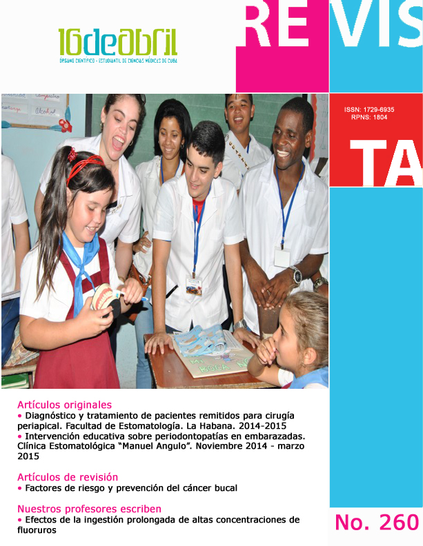
Diagnóstico y tratamiento de pacientes remitidos para cirugía periapical. Facultad de Estomatología. La Habana, 2014-2015
Palabras clave:
procedimientos quirúrgicos orales, evaluación preoperatoria, salud bucal, oral surgical procedures, surgical clearance, oral healthResumen
Introducción: El estado periapical y la calidad de la terapia endodóntica son evaluados para la cirugía periapical. Objetivo: Describir la conducta diagnóstica y terapéutica ante pacientes remitidos para cirugía periapical. Materiales y Métodos: Se realizó un estudio observacional, descriptivo, transversal, desde enero 2014 a enero 2015, en la Facultad de Estomatología ¨Raúl González Sánchez¨. Se trabajó con 57 pacientes remitidos al Departamento de Cirugía Maxilofacial para cirugía periapical. Resultados: La causa más frecuente de remisión fue la presencia de área radiolúcida periapical ≤ 1cm (47,37%). El error diagnóstico más identificado fue la remisión de pacientes con áreas radiolúcidas sin tratamiento pulporadicular realizado (28,07%). El error terapéutico más detectado fue la obturación ductal con defecto de extensión (38,60%). El (29,82%) de los pacientes fue tratado mediante curetaje apical. Conclusiones: El principal error diagnóstico fue la remisión de áreas radiolúcidas sin tratamiento pulpo radicular. El error terapéutico principal fue el defecto de extensión de la obturación ductal.
ABSTRACT
Introduction: The periapical status and quality of endodontic therapy are evaluated for periapical surgery. Objective: To describe the diagnostic and therapeutic approach to patients referred for periapical surgery. Materials and Methods: An observational, descriptive, cross-sectional study was conducted from January 2014 to January 2015, at the Faculty of Stomatology Raúl González Sánchez. We worked with 57 patients referred to the Department of Maxillofacial Surgery for periapical surgery. Results: The most frequent cause of referral was the presence of periapical radiolucent area ≤1 cm (47.37%). The misdiagnosis was identified referral of patients with radiolucent areas without pulporadicular treatment performed (28.07%). The mistake was detected therapeutic shutter ductal extension defect (38.60%). The (29.82%) patients was treated by apical curettage. Conclusions: The main misdiagnosis was remission of radiolucent areas untreated root octopus. The main therapeutic mistake was the fault of the ductal extension seal.
Citas
2. Rodríguez R, Torres D, Gutiérrez JL. ¨Puesta al día en cirugía endodóntica¨. Sociedad española de Cirugía Bucal. Rev SECIB On Line 2008; V1: pp1 – 15.
3. Persic R, Kqiku L. Difference in the periapical status of endodontically treated teeth between the samples of Croatian and Austrian adult patients. Croat Med J. 2011; 52(6): 672–678.
4. Ureyen K, Kececi A. A retrospective radiographic study of coronal periapical status and root canal filling quality in a selected adults. Med Princ Pract. 2013; 22(4):334-9.
5. Sherwood A. Pre-operative diagnostic radiograph interpretation by general dental practitioners for root canal treatment. Dentomaxillofac Radiol. 2012; 41(1): 43–54.
6. Matijevic J, Cizmekovic T. Prevalence of apical periodontitis and quality of root canal fillings in population of Zagreb, Croatia: a cross-sectional study. Croat Med J. 2011; 52(6): 679–687.
7. Chala S, Abougal R. Prevalence of apical periodontitis and factors associated with the periradicular status. Acta Odontol Scand. 2011; 69(6):355-359.
8. Tavares P, Bonte E. Prevalence of Apical Periodontitis in Root Canal–Treated Teeth From an Urban French Population: Influence of the Quality of Root Canal Fillings and Coronal Restorations. Journal of Endodontics. 2009;35(6):810-813.
9. Fernández M, Ataide I. Nonsurgical management of periapical lesions. J Conserv Dent.2010; 13(4): 240–245.
10. Dandotikar D. Peddi R. Non Surgical management of a Periapical Cyst. : A case report. J Int OraL Health. 2013; 5(3):79-84.
11. Ertas E, Ertas H. Radiographic Assessment of the Technical Quality and Periapical Health of Root-Filled Teeth Performed by General Practitioners in a Turkish Subpopulation. ScientificWorldJournal. 2013; 2013: 514841. Available from http://dx.doi.org/10.1155%2F2013%2F514841
12. Mukhaimer R. Prevalence of apical periodontitis and quality of root canal treatment in an adult Palestinian sub-population. Saudi Dent J. 2012; 24(3-4): 149–155.
13. Von Arx T. Apical surgery: A review of current techniques and outcome. Saudi Dent J. 2011; 23(1): 9–15.
14. European Society of Endodontology. Quality Guidelines of endodontic treatment: consensus report. Int Endod J. 2006; 39(12):921-30.
15. Ricucci D, Russo j. A prospective cohort study of endodontic treatments of 1,369 root canals: results after 5 years. Oral Surg. Oral Med. Oral Pathol. Oral Radiol. Endod. 2011 Dec [cited 2014 Feb 17];112(6):825-842.
16. Marti E, Peñarrocha M. An update in periapical surgery. Med. oral patol. oral cir.bucal.2006; 11(6):6-7.
17. Rahbaran S, Gilthorpe M. Comparison of clinical outcome of periapical surgery in endodontic and oral surgery units of a teaching dental hospital: A retrospective study. Oral Surg. Oral Med. Oral Pathol. Oral Radiol. Endod. 2001;91(6):700-709.
18. Garcia B, Martorel L. Cirugía periapical en dientes posteriores maxilares. Revisión de la bibliografía Periapical surgery of maxillary posterior teeth. A review of the literature. Med. oral patol. oral cir.bucal. 2006; 11(2):45-47.
19. Peñarrocha M, Gay Escoda C. A prospective clinical study of polycarboxylate cement in periapical surgery. Med Oral Patol Oral Cir Bucal.2012; 17(2):276–280.
20. Walivaara D, Abrahamson P. Super-EBA and IRM as root-end fillings in periapical surgery with ultrasonic preparation: a prospective randomized clinical study of 206 consecutive teeth. Oral Surg. Oral Med. Oral Pathol. Oral Radiol. Endod. 2011;112(2):258-263.
21. Vallecillo M, Muñoz E, Reyes C, Prados E, Olmedo MªV. Cirugía periapical de 29 dientes. Comparación entre técnica convencional, microsierra y uso deultrasonidos. Medicina Oral 2002; 7: 46-53. Disponible en: http://www.medicinaoral.com/pubmed/medoralv7_i1_p46.pdf
22. Lingyung S, Yang G. Surgical endodontic treatment of refractory periapical periodontitis with extraradicular biofilm. . Oral Surg. Oral Med. Oral Pathol. Oral Radiol. Endod. 2010;110(1):40-44.
23. Asgary S, Shadman B. Periapical Status and Quality of Root canal Fillings and Coronal Restorations in Iranian Population. Iran Endod J. 2010; 5(2): 74–82.
24. Gencoglu N. Periapical Status and Quality of Root Fillings and Coronal Restorations in an Adult Turkish Subpopulation. Eur J Dent. 2010 Jan; 4(1): 17–22.
25. Moreno J, Alves F. Periradicular Status and Quality of Root Canal Fillings and Coronal Restorations in an Urban Colombian Population. Journal of Endodontics. 2013;39(5):600-604.
26. Kim S. Prevalence of apical periodontitis of root canal–treated teeth and retrospective evaluation of symptom-related prognostic factors in an urban South Korean population. . Oral Surg. Oral Med. Oral Pathol. Oral Radiol. Endod. 2010; 110(6): 795–799.
27. Kim S, Kratchman S. ¨Modern endodontic surgery concepts and practice¨. Endodont . J. 2006; 32: 601-623.
28. Del Fabbro M, Taschieri S, Testori T, Francetti L, Weinstein RL. Nuevo tratamiento endodóncico quirúrgico versus no quirúrgico para las lesiones perirradiculares¨ octubre 8, 2008. Disponible en: http://summaries.cochrane.org/es/CD005511/nuevo-tratamiento-endodoncico-quirurgico-versus-no-quirurgico-para-las-lesiones-perirradicula
Descargas
Publicado
Cómo citar
Número
Sección
Licencia
Aquellos autores/as que tengan publicaciones con esta revista, aceptan los términos siguientes:
Los autores/as conservarán sus derechos de autor y garantizarán a la revista el derecho de primera publicación de su obra, el cuál estará bajo una Licencia de Creative Commons Reconocimiento-NoComercial 4.0 Internacional (CC BY-NC 4.0)
Los autores/as podrán adoptar otros acuerdos de licencia no exclusiva de distribución de la versión de la obra publicada (p. ej.: depositarla en un archivo telemático institucional o servidores preprint) siempre que se indique la publicación inicial en esta revista.
Se permite y recomienda a los autores/as difundir su obra a través de Internet (p. ej.: en archivos telemáticos institucionales o en su página web) antes y durante el proceso de envío, lo cual puede producir intercambios interesantes y aumentar las citas de la obra publicada.


